Bipolar Neuron: Definition, Structure, Location, Function, and Examples
In the intricate landscape of the human nervous system, neurons are the fundamental building blocks responsible for the transmission of electrical and chemical signals. While most people are familiar with the classic “tree-like” multipolar neuron often depicted in textbooks, the nervous system utilizes a variety of specialized shapes to perform specific tasks. Among the most specialized and functionally distinct is the bipolar neuron.
Understanding what a bipolar neuron is essential for students of biology, nursing, and medicine, as these cells play a pivotal role in our sensory perception. This article provides a deep dive into the definition, anatomical structure, histological appearance, and vital functions of bipolar neurons, while offering a clear comparison to other neuronal types.
What Is a Bipolar Neuron?
At its most fundamental level, a bipolar neuron is a type of nerve cell characterized by having exactly two processes (projections) extending from opposite ends of the cell body (soma). The term “bipolar” literally translates to “two poles,” referring to these two distinct extensions.
In the vast majority of neurons in the human body, such as those found in the brain and spinal cord, there are many dendrites and one axon (multipolar). However, in a bipolar neuron, the design is simplified and highly linear. One process serves as a dendrite, receiving sensory information from the environment or a receptor cell, while the other process serves as the axon, transmitting that signal toward the Central Nervous System (CNS).
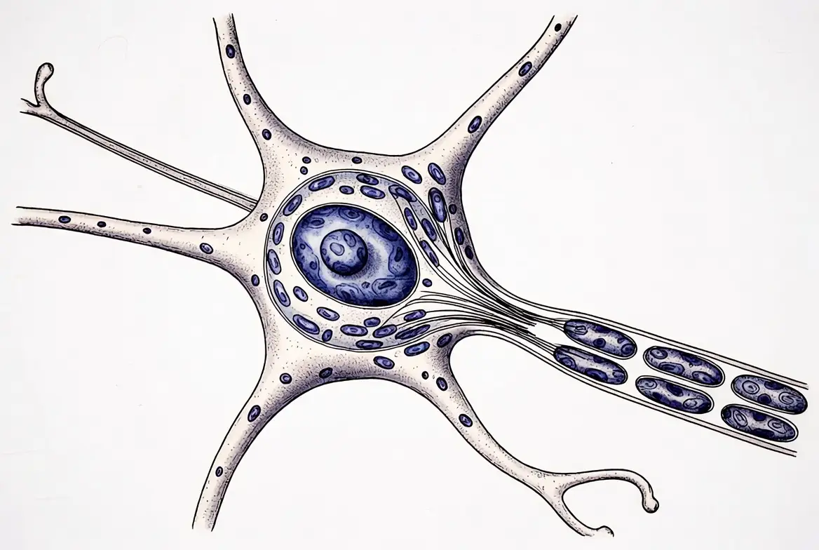
Structure of a Bipolar Neuron
When asking what a bipolar neuron looks like, the answer is simple. The bipolar neuron structure is remarkably symmetrical compared to its multipolar cousins.
Cell Body (Soma)
The soma of a bipolar neuron is typically spindle-shaped (fusiform) or oval. It contains the nucleus and the necessary organelles for cellular maintenance, such as the mitochondria and Golgi apparatus. Unlike multipolar neurons, where the soma is covered in dendrites, the soma of a bipolar neuron is relatively “clean,” with only two exit points.
One Dendrite + One Axon
- The Dendrite: This process extends toward the periphery. It often branches into a small dendritic arbor at its tip to receive input from sensory receptors.
- The Axon: This process extends toward the CNS. It conducts the action potential away from the cell body.
Direction of Signal Flow
The signal flow in a bipolar neuron is strictly unidirectional. It enters through the distal end of the dendrite, passes through the cell body, and travels down the axon to the synaptic terminals.
Bipolar Neuron Diagram and Labeled Anatomy
Identifying these cells in a lab or on a test requires a clear understanding of a bipolar neuron diagram. A standard bipolar neuron labeled diagram will include the following parts:
- Dendrite: The process of receiving information.
- Dendritic Branches: Terminal ends that synapse with receptor cells.
- Soma (Cell Body): The central hub containing the nucleus.
- Nucleus: The genetic heart of the cell.
- Axon: The long process conducting the impulse.
- Axon Terminals: The end of the neuron that releases neurotransmitters.
Exam Labeling Tips
In many diagrams, the dendrite and axon look nearly identical. To tell them apart, look for the direction of the signal (if indicated) or look for the receptor cells. The process attached to the receptor (like a rod or cone cell in the retina) is the dendrite.
What Does a Bipolar Neuron Look Like?
Visually, the bipolar neuron is defined by its linear shape. While multipolar neurons look like exploded stars or trees, the bipolar neuron looks more like a bridge. It is often described as being “fusiform,” meaning it tapers at both ends.
Comparison to Unipolar and Multipolar Neurons
- Multipolar neurons have a large, star-shaped soma with many branching dendrites.
- Unipolar neurons have a soma with only a single process that eventually splits.
- Bipolar neurons are unique because the soma sits directly in the middle of a straight line formed by the axon and dendrite.
This appearance is a direct reflection of its function: it is not meant to integrate a thousand different signals at once; it is designed to be a high-fidelity, one-to-one relay.
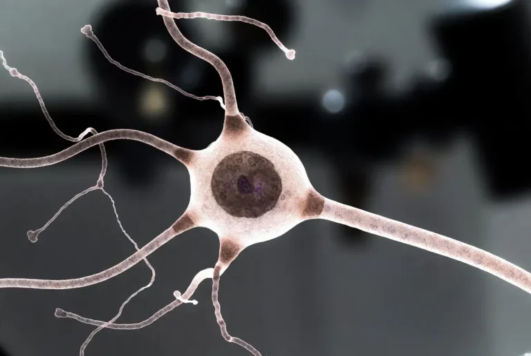
Bipolar Neuron Under Microscope
In a laboratory setting, observing a bipolar neuron under a microscope is a rare treat because of its limited location in the body. Typically, students will view these through prepared slides of the retina or olfactory epithelium.
Light Microscopy Appearance
Under standard light microscopy with a Golgi stain (which turns neurons black or dark brown), a bipolar neuron appears as a dark, oval center with two thin lines extending from it.
Histological Staining Methods
- H&E Stain: Difficult to identify individual processes; usually, only the soma is clearly visible.
- Silver Staining (Golgi’s Method): The gold standard for seeing the full architecture of a bipolar neuron.
- Fluorescent Labeling: Used in modern research to track specific bipolar cells in the retina.
Common Lab Observations
When performing a bipolar neuron microscopy observation, you will notice that these cells are often packed tightly together. In the retina, for example, they form a distinct “Bipolar Cell Layer” between the photoreceptors and the ganglion cells.
Bipolar Neuron Histology
The study of bipolar neuron histology involves identifying these cells within their native tissues. Because they are part of sensory pathways, they are always surrounded by supporting cells (glia) and other neurons.
In a tissue section of the retina, you can identify the bipolar layer by looking for the middle layer of nuclei. Unlike the “pseudo-unipolar” neurons found in the dorsal root ganglia (which have large, round cell bodies), retinal bipolar neurons are smaller and more elongated.
Function of a Bipolar Neuron
The primary function of a bipolar neuron is sensory transmission. They are the “middlemen” of the special senses. They do not usually act as the initial receptor, nor do they carry the signal all the way to the primary processing centers of the brain; instead, they act as a vital bridge.
Role in Sensory Signal Transmission
A bipolar neuron does the job of condensing and passing on information. For example, in the eye:
- Rods and Cones (Photoreceptors) detect light.
- The Bipolar Neuron receives this chemical signal.
- The Bipolar Neuron passes the signal to Ganglion Cells, whose axons form the optic nerve.
Signal Flow from Receptor to CNS
The function of bipolar neuron pathways is to ensure that the “raw data” from the environment is translated into a neural code that the brain can eventually interpret as an image, a smell, or a sound.
Is a Bipolar Neuron a Sensory Neuron?
Yes. Every bipolar neuron in the human body is a sensory neuron. However, the inverse is not true: not every sensory neuron is bipolar.
- Motor Neurons: Are almost exclusively multipolar.
- Interneurons are almost exclusively multipolar.
- Sensory Neurons: Can be unipolar (pseudo-unipolar) or bipolar.
Bipolar neurons are specialized for the special senses (vision, olfaction, hearing, and balance), while the general senses (touch, pain, and temperature) utilize pseudo-unipolar neurons. This distinction is a favorite topic for anatomy and physiology instructors.
Location of Bipolar Neurons in the Body
One of the most common exam questions is: Where is the bipolar neuron located? Unlike multipolar neurons, which are found everywhere, the location of bipolar neuron cells is highly restricted to specific sensory organs.
Bipolar Neuron Location — CNS or PNS?
This is a complex question. Most bipolar neurons are considered part of the Peripheral Nervous System (PNS) because they are associated with sensory receptors outside the brain and spinal cord. However, the retina is embryologically an outgrowth of the brain, leading many to argue that retinal bipolar cells are technically part of the CNS.
Specific Locations:
- The Retina (Eye): Found in the intermediate layer of the retina.
- Olfactory Epithelium (Nose): Used to transmit the sense of smell.
- Vestibulocochlear Nerve (Ear): Found in the spiral ganglion (hearing) and vestibular ganglion (balance).
Examples of Bipolar Neurons
To fully grasp what an example of a bipolar neuron is, one must look at the specific sensory systems where these cells are indispensable. Because they are so rare, they only appear in highly specialized anatomical “niches.”
Retina Bipolar Cells
The most famous bipolar neuron example is found in the eye. In the retina, these cells act as a high-speed conduit. There are several types of retinal bipolar cells, including “on-center” and “off-center” cells, which help the brain detect contrast and edges in a visual field. Without these bipolar neurons, the information captured by your rods and cones would never reach the optic nerve.
Olfactory Receptor Neurons
In the upper reaches of the nasal cavity lies the olfactory epithelium. Here, bipolar neurons serve as the actual receptors for smell. The dendrites of these neurons have specialized cilia that bind to odorant molecules. When a smell is detected, the bipolar neuron sends a signal directly through the cribriform plate of the ethmoid bone and into the olfactory bulb of the brain.
Vestibular and Cochlear Neurons
In the inner ear, bipolar neurons are responsible for transmitting two distinct types of data:
- Cochlear Neurons: Located in the spiral ganglion, these relay auditory (sound) signals.
- Vestibular Neurons: Located in the vestibular ganglion (Scarpa’s ganglion), these relay information regarding balance, head position, and spatial orientation.
Bipolar Neuron Abundance — Why Are They Rare?
In the human body, multipolar neurons make up about 99% of all nerve cells. This raises the question: Why is the bipolar neuron abundance so low?
The answer lies in functional specialization. Most tasks in the nervous system—such as thinking, planning, and moving muscles—require the integration of thousands of inputs. A motor neuron needs to “listen” to many different brain regions before deciding to contract a muscle, which is why it has many dendrites (multipolar).
Bipolar neurons, however, are designed for fidelity. In the sensory systems, you don’t want the signal to be “mixed” or diluted by other inputs. You want the light hitting a specific part of the retina to be reported as accurately as possible. The “one-to-one” structure of the bipolar neuron is an evolutionary solution for precise, fast sensory relay.
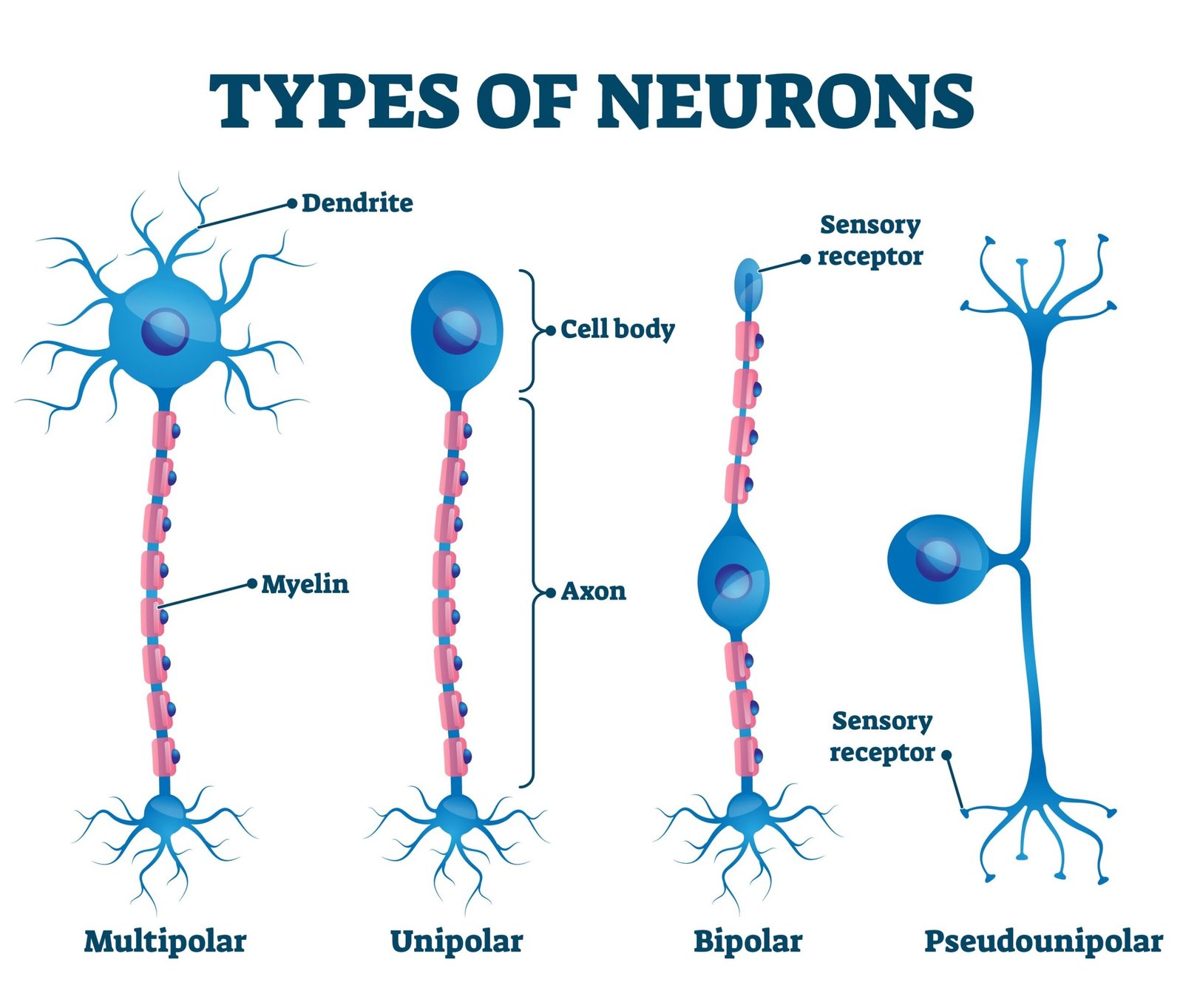
Types of Neurons — Unipolar, Bipolar, and Multipolar
To understand what type of neuron a bipolar neuron is, it is helpful to place it within the broader classification of the nervous system. Scientists categorize neurons based on their morphology (shape).
- Unipolar (Pseudo-unipolar) Neurons: These have a single process that extends from the cell body and then splits into two branches. One branch goes to the periphery (sensory receptor) and the other goes to the spinal cord. These are the most common sensory neurons for touch and pain.
- Bipolar Neurons: As we have explored, these have two distinct processes. They are exclusively sensory and rare.
- Multipolar Neurons: These are the “standard” neurons with one axon and many dendrites. They include motor neurons and interneurons.
Bipolar Neuron vs. Unipolar Neuron
The difference between a bipolar and unipolar neuron is a frequent source of confusion for students, primarily because both are involved in sensory pathways.
| Feature | Bipolar Neuron | Unipolar (Pseudo-unipolar) Neuron |
| Structure | Two processes from opposite ends of the soma. | One process that splits into a “T” shape. |
| Soma Location | Centrally located between the processes. | Offset to the side of the main axon. |
| Soma Shape | Elongated or spindle-shaped. | Typically round. |
| Primary Location | Retina, Olfactory, Inner Ear (Special Senses). | Dorsal Root Ganglia (General Senses like touch/pain). |
| Function | Specialized relay for special senses. | General sensory conduction to the spinal cord. |
Bipolar Neuron vs. Multipolar Neuron
When comparing bipolar vs multipolar neurons, the distinction is even more stark. A multipolar neuron is built for complexity and “data processing,” while a bipolar neuron is built for “data transport.”
- Dendritic Count: A bipolar neuron has exactly one dendrite. A multipolar neuron can have hundreds or thousands.
- Synaptic Integration: Multipolar neurons perform “summation,” where they add up all the signals they receive to decide whether to fire. Bipolar neurons generally pass the signal along with very little modification.
- Prevalence: Multipolar neurons are the most common; bipolar neurons are among the rarest.
Pseudo-Unipolar Neurons Explained
For years, many textbooks simply referred to sensory neurons in the spinal cord as “unipolar.” However, modern histology uses the term pseudo-unipolar neuron.
These neurons start as bipolar neurons during embryonic development. As the fetus grows, the two processes fuse together at the base, resulting in a single stalk that emerges from the cell body. This stalk then bifurcates (splits) into a peripheral and a central branch. While they look like they have only one process, they are functionally different from the true unipolar neurons found in invertebrates.
Bipolar Neurons and Nerve Pathways
Bipolar nerves (nerves composed largely of bipolar axons) are essential for the “Special Visceral” and “Special Somatic” afferent pathways.
These pathways usually involve a chain of three neurons to get a signal from the receptor to the conscious part of the brain (the cerebral cortex). These are known as First-Order, Second-Order, and Third-Order Neurons.
What Is a 1st, 2nd, and 3rd Order Neuron?
- 1st Order Neuron: This is the neuron that first detects the stimulus. In the olfactory and inner ear systems, the bipolar neuron is the 1st order neuron. (In the retina, the photoreceptor is technically the 1st order cell, and the bipolar cell is the 2nd order cell.
- 2nd Order Neuron: This neuron carries the signal to the thalamus, which is the “relay station” of the brain.
- 3rd Order Neuron: This neuron carries the signal from the thalamus to the sensory cortex for conscious perception.
Frequently Asked Questions
What is a bipolar neuron?
A bipolar neuron is a type of nerve cell with two extensions—one axon and one dendrite—coming off a central cell body.
Where is the bipolar neuron located?
They are found in the specialized sensory organs: the retina of the eye, the olfactory epithelium of the nose, and the auditory/vestibular organs of the inner ear.
How many dendrites does a bipolar neuron have?
A bipolar neuron has exactly one dendrite.
Is a bipolar neuron sensory or motor?
It is always sensory. It carries information from receptors toward the central nervous system.
Why are bipolar neurons rare?
Because they are highly specialized for the high-fidelity transmission of special senses. Most of the body’s tasks require the complex signal integration provided by multipolar neurons.
Conclusion
Though they represent only a tiny fraction of the billions of neurons in the human body, bipolar neurons are the gatekeepers of our reality. They are the essential links that allow the chemical signature of a rose, the light from a distant star, or the melody of a song to be translated into the language of the brain.
For the student, mastering the structure and function of a bipolar neuron is more than just memorizing a definition for an exam; it is an exploration of how evolution has crafted a perfect “bridge” for our most precious senses. By understanding how these cells differ from unipolar and multipolar neurons, we gain a deeper appreciation for the elegant specialization of the human nervous system.
Authoritative References
1. National Center for Biotechnology Information (NCBI) – StatPearls: Histology, Central Nervous System
2. Kenhub – Types of Neurons: Structure and Functions
3. Molecular Biology of the Cell (NCBI Bookshelf) – The Retina
4. LibreTexts Biology – Neurons and Glial Cells
5. Gray’s Anatomy – The Organs of the Senses

Related Posts


Subscribe to Our Newsletter
Get mental health tips, updates, and resources delivered to your inbox.
MORE from Author
Read More In my practice as a clinical psychologist, I often observe that the difference between a patient who crumbles under pressure…
- We all have "up" days and "down" days. Perhaps you feel energized after a promotion or sluggish on a rainy…


 To define bipolar disorder accurately, we must look at it as a chronic mood dysregulation condition. In psychology and medicine,…
 To answer whether bipolar fits the bill, we first have to define what a neurodivergent person actually is. The term…


Are you looking for a Therapist?
Connect with qualified mental health professionals who understand
bipolar disorder, mood changes, and emotional challenges.
Private • Supportive • Confidential